Advanced Minimally Invasive Diagnosis for Lung and Thoracic Diseases
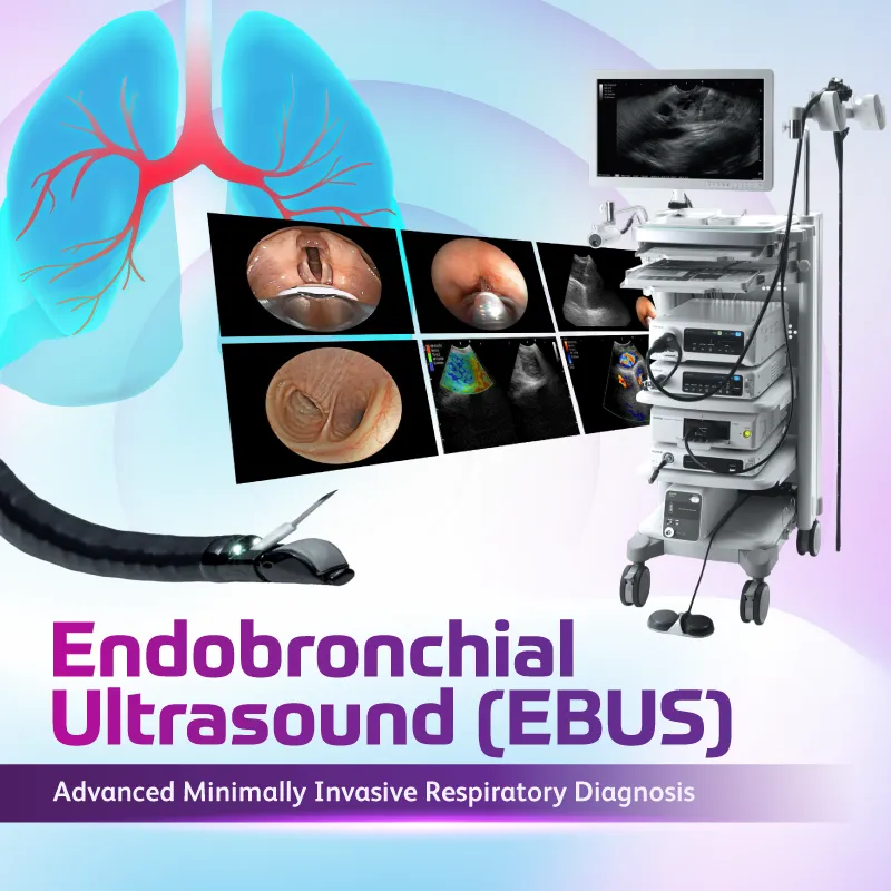
Endobronchial Ultrasound (EBUS) is an advanced diagnostic procedure that combines bronchoscopy with ultrasound imaging to examine the lungs, airways, and surrounding structures within the chest.
This minimally invasive technology allows doctors to obtain real-time ultrasound images from inside the airways, enabling them to detect abnormalities, check lymph nodes and perform targeted biopsies with high precision.
EBUS plays an important role in the early detection, diagnosis, and staging of lung cancer, as well as in the evaluation of other respiratory and thoracic conditions.
What is Endobronchial Ultrasound (EBUS)?
During an EBUS procedure, a thin and flexible bronchoscope equipped with a miniature ultrasound probe is inserted through the mouth or nose into the airways.
The ultrasound probe produces high-resolution images of structures beyond the airway walls, allowing doctors to examine areas that cannot be seen with conventional bronchoscopy alone.
Watch Our Video :
Learn More About EBUS: A Safer Way to Diagnose and Stage Lung Cancer by Dr. Wan Jen Lye
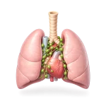
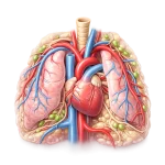
EBUS enables detailed evaluation of important thoracic structures, including:
Lungs
Trachea and bronchi (airways)
Mediastinum (the area between the lungs)
Mediastinal lymph nodes
Nearby blood vessels and tissues
By visualising these structures in real time, doctors can identify abnormalities and collect tissue samples safely and accurately.
Why EBUS is Important in Lung Cancer Diagnosis
EBUS has become an essential tool in modern thoracic and respiratory medicine, particularly for patients suspected of having lung cancer
One of its key roles is lung cancer staging, which determines whether cancer has spread to nearby lymph nodes in the chest. Accurate staging is critical for deciding the most appropriate treatment approach, especially in early stage lung cancer where curative surgery is a treatment option.
With real-time ultrasound guidance, EBUS allows doctors to perform Transbronchial Needle Aspiration (TBNA), a technique where a fine needle is used to collect tissue samples to obtain tissue samples from suspicious lymph nodes or lung lesions without the need for surgical procedures.
This improves diagnostic accuracy while reducing the risks and recovery time associated with more invasive techniques.
Types of EBUS
There are two main types of EBUS used depending on the location of the lung abnormality.
Linear EBUS
Linear EBUS is commonly used to examine and sample mediastinal lymph nodes and central lung lesions. It allows doctors to perform real-time needle biopsies and is widely used for lung cancer diagnosis and staging.
Radial EBUS
Radial EBUS uses a miniature rotating ultrasound probe to evaluate peripheral lung lesions, including small lung nodules located deeper within the lungs. This technique helps doctors access areas that are difficult to reach with standard bronchoscopy.
Benefits of Endobronchial Ultrasound (EBUS)
EBUS offers several advantages compared to conventional diagnostic methods.
Minimally Invasive
The procedure is typically performed under local anaesthesia with sedation, without the need for major surgery.
Supports Early Detection of Lung Disease
EBUS can detect small lung nodules or lymph node abnormalities that may not be visible with standard imaging or bronchoscopy.
Accurate Lung Cancer Staging
It enables doctors to determine whether lung cancer has spread to nearby lymph nodes, which is essential for treatment planning.
Precise Guided Biopsy
Using real-time ultrasound imaging, doctors can obtain targeted tissue samples from difficult-to-reach areas safely.
Lower Risk of Complications
Compared to surgical biopsies, EBUS has a lower risk of bleeding and pneumothorax.
Faster Recovery
Patients usually recover quickly and may be discharged within 1–2 days if stable
Conditions That Can Be Diagnosed with EBUS
EBUS can help diagnose and evaluate a wide range of lung and thoracic conditions, including:
Lung cancer
Enlarged mediastinal lymph nodes
Pulmonary nodules or masses
Lung infections
Inflammatory lung diseases
Other thoracic abnormalities
Early and accurate diagnosis allows doctors to plan timely and appropriate treatment for better patient outcomes.
What Happens During an EBUS Procedure?
1
Preparation
Patients are required to fast for at least 6 hours before
the procedure.
2
Sedation
Intravenous sedation or local anaesthesia is administered
while vital signs are monitored.
3
Bronchoscope Insertion
A specialised ultrasound bronchoscope is gently inserted
through the mouth or nose into the airways.
4
Ultrasound Imaging
The ultrasound probe provides real-time images of the lungs,
lymph nodes, and surrounding structures.
5
Tissue Sampling (TBNA)
A fine needle is passed through the bronchoscope to obtain
tissue samples from suspicious areas. The collected samples
are prepared for immediate examination to ensure sufficient
tissue is obtained for further diagnostic evaluation. This
process also helps improve the accuracy of the procedure by
ensuring the correct tissue is obtained for further analysis.
6
Recovery
After the procedure, patients are monitored until the sedation
wears off. Patients should arrange for someone to accompany
them home, as drowsiness may persist for a few hours.
Frequently Asked Questions (FAQ)
Most patients receive sedation, so discomfort is minimal. A mild sore throat or cough may occur after the procedure.
The procedure typically takes 30 to 60 minutes, depending on the areas examined and whether biopsies are performed.
EBUS is generally safe with a low complication rate. Minor risks may include bleeding, infection, mediastinitis or reactions to sedation.
Preliminary findings may be discussed with doctor shortly after the procedure, while biopsy results are usually available within a few days.
Many patients can go home on the same day or within 1–2 days, depending on their condition.
Meet Our Specialists
Dr Goh Kee San
Consultant Respiratory Medicine & Internal Medicine Physician