紧急热线: +603 7620 7979 / +603 7787 2992


患者需在检查前至少禁食8小时。
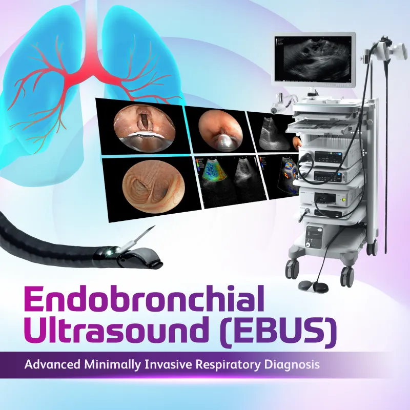
大多数患者会接受镇静,因此不适感较轻。检查后可能会出现轻微喉咙痛或咳嗽。
检查通常需要30至60分钟,具体时间取决于检查范围及是否进行活检。
EBUS总体上是安全的,并发症发生率较低。轻微风险包括出血、感染、纵隔炎或对镇静药物的反应。
初步结果可能在检查后不久即可讨论,而活检结果通常需数天后才能获得。
多数患者可于当天或1至2天内出院,具体视病情而定。
Monday – Friday
8:30 AM – 6:00 PM