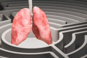
In respiratory medicine, one of the most difficult challenges is not always the treatment, but getting to the right diagnosis early, safely, and accurately. Diseases within the chest can be hidden deep within, not easily accessible, and for many years, this meant that reaching a diagnosis may require trials and sometimes more invasive procedures.
Endobronchial Ultrasound, or EBUS, has changed this landscape in a significant way. It represents an upgrade in diagnostic applications—allowing clinicians to see beyond the airway wall, obtain targeted tissue samples, and reduce the need for surgical exploration in many cases.
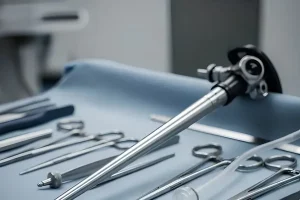
EBUS combines two established techniques: bronchoscopy and ultrasound imaging. A thin, flexible bronchoscope is gently inserted through into the airways, similar to bronchoscopy. What makes EBUS different is the addition of ultrasound that allows us to visualise structural areas that were previously difficult to assess without surgery. From a clinical perspective, instead of relying on indirect imaging alone, EBUS allows direct, real-time visualisation of areas from within the airway itself.
One of the notable applications of the EBUS is in lung cancer diagnosis and staging. In lung cancer care, staging determines the extent of disease spread and directly influences treatment decisions. Lymph nodes found in the chest play a critical role in this assessment. Traditionally, accessing these lymph nodes often required surgical procedures such as mediastinoscopy, which, while effective, are more invasive and require general anaesthesia.
With EBUS, we are able to evaluate these lymph nodes in real-time, plus, the ultrasound-guided biopsies using fine needles attached to the bronchoscope, we can obtain tissue samples for diagnosis and staging under the same procedure. This approach significantly improves diagnostic precision while reducing procedural risk and patient discomfort.
Beyond lung cancer, EBUS also has an important role in the examination of other thoracic conditions. These include unexplained enlarged lymph nodes, inflammatory conditions such as sarcoidosis, and infections that affect the mediastinum or lymphatic structures. In these scenarios, EBUS provides a minimally invasive way towards biopsy before initiating long-term treatment.

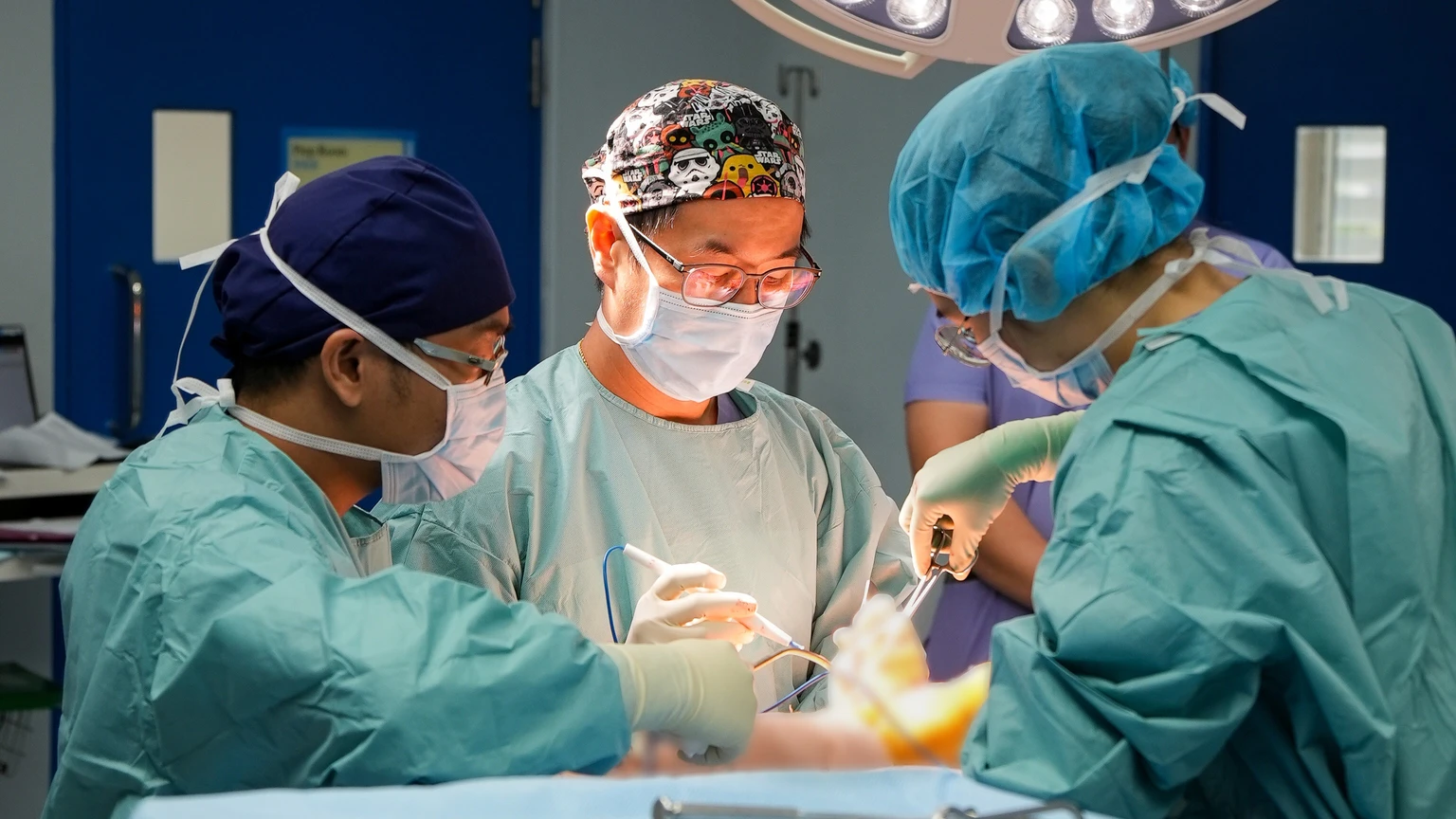
Aside from the technical capability of the procedure, the EBUS can profoundly impact on patient care. Being able to reach a diagnosis with less physical toll on the patient could potentially change the receptivity towards the procedure. This minimally invasive procedure requires sedation instead of anaesthesia, and most patients are more comfortable knowing they can recover faster compared to surgery.
When putting EBUS into perspective, it is not just about technological advancement, but rather minimising the burden while improving diagnostic clarity. Progress in medicine is not always about adding more variety, but about doing things more precisely. And in the complexity and critical structure of the chest, precision makes a meaningful difference.
Dr Wan Jen Lye